Diabetic Eye Disease and Vitreoretinal Surgery: Navigating Treatment Options

Diabetic eye disease like diabetic retinopathy can permanently alter a person’s vision, leading to vision loss or, in severe cases, blindness. With diabetic retinopathy, the blood vessels in the eye weaken. At times, that can lead vessels to bulge and leak, and the latter can cause fluids to enter various parts of the eye, causing retinal tissue swelling. As those tissues are impacted, visual acuity changes, and any damage caused is potentially irreversible.  

However, treating diabetic eye disease can slow or prevent future eye changes, allowing a person to limit their vision loss. One such option is vitreoretinal surgery, also known as vitrectomy, but different paths are also available.  

Here’s a look at vitreoretinal surgery and a few other treatments for diabetic eye disease, as well as what you can do to navigate your treatment options.  

Vitreoretinal Surgery for Diabetic Retinopathy 

Vitreoretinal surgery is a procedure that’s often used in the treatment of advanced diabetic retinopathy. While it isn’t a cure for diabetic retinopathy, it can slow its progression and may reverse some of its impact, causing your vision to possibly improve, stabilize, or avoid significant ongoing losses.  

With a vitrectomy, your eye care provider addresses vitreous gel clouding that can occur when blood vessels leak by removing impacted tissue and replacing it with a clear artificial substitute. In many cases, replacing the cloudy vitreous gel with a clear artificial solution leads to significant vision improvement.  

During the procedure, your eye care provider will also remove built-up scar tissue that’s developed in the eye. Scar tissue can pull on the retina, leading to visual distortions and increasing the chances of a retinal detachment. As a result, scar tissue removal can lead to vision improvements and reduce the risk of retinal detachment.  

Other Treatments for Diabetic Retinopathy

Generally, vitreoretinal surgery is reserved for advanced diabetic retinopathy cases. Other treatment options may provide positive results if your case isn’t that severe. For example, blood sugar management could prevent further damage, which may make additional treatment unnecessary.  

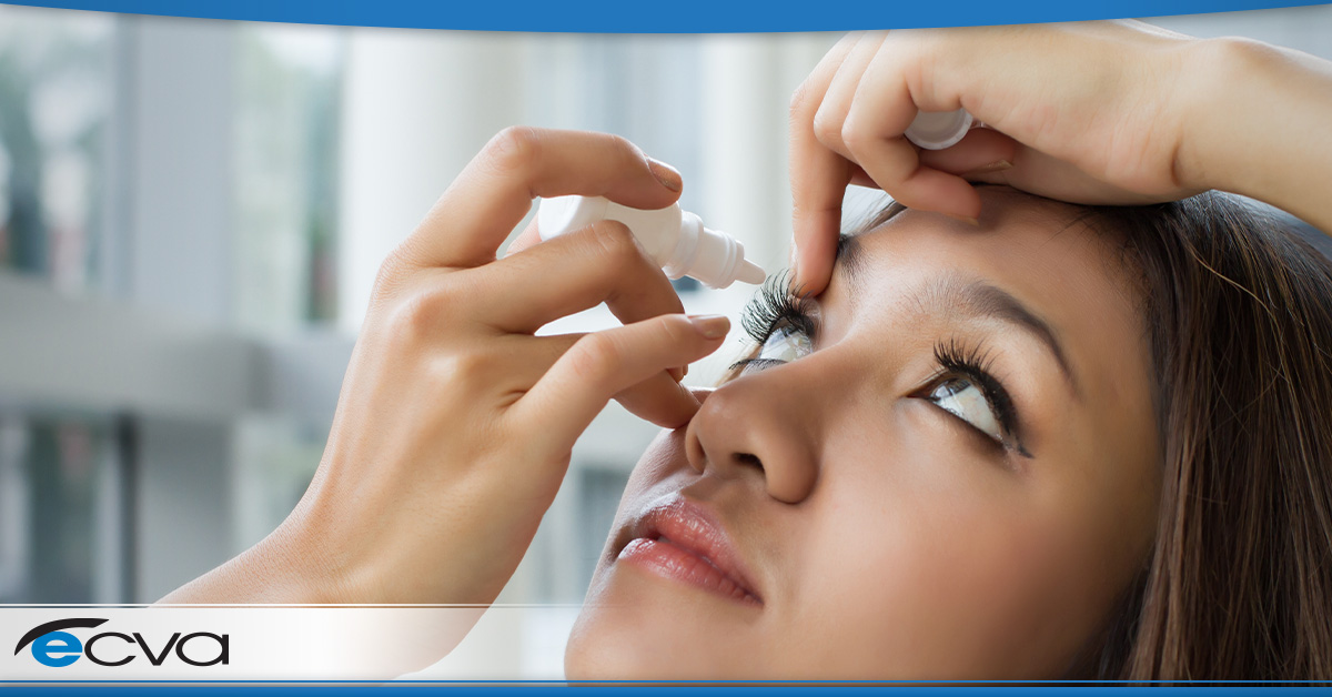
For more severe cases, medication injections involving vascular endothelial growth factor inhibitors can help prevent abnormal blood vessel development that can occur with diabetic retinopathy and reduce fluid buildup. Focal laser or scatter laser treatment – also known as photocoagulation – can also slow or stop blood vessel leakage or shrink abnormal vessels, making them a viable option in some cases.  

How to Navigate Your Treatment Options

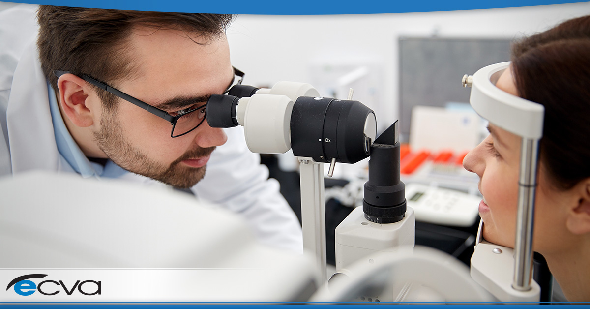
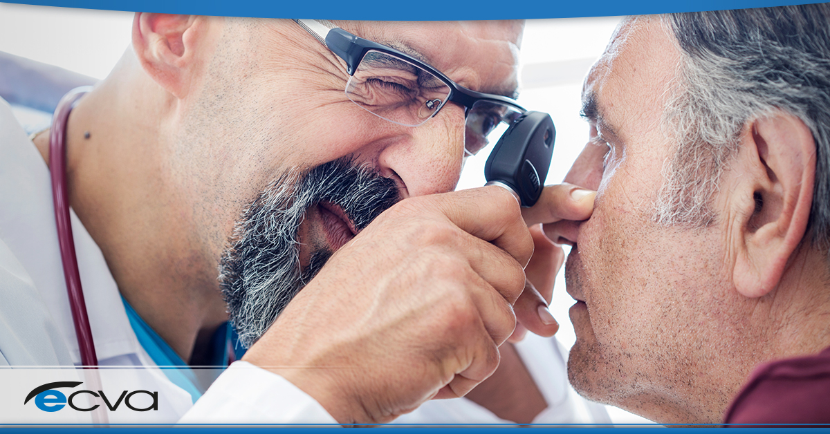
Selecting a treatment option after being diagnosed with diabetic retinopathy is often intimidating. Fortunately, by working with your eye care provider, you can receive critical guidance and support that makes selecting an appropriate approach easier. 

Your eye care provider can look at the details of your situation, identifying treatment options that are both viable and have the highest chances of making a positive impact. They can also answer questions to help you understand the recommendations, ensuring you can find the best treatment option for you.  

At ECVA, the safety and health of our patients’ eyes are our priority. If you’ve been diagnosed with diabetic retinopathy, are concerned you may develop diabetic eye disease, or simply haven’t visited your eye care provider in the past year, the ECVA team is here to help. Schedule an appointment at your closest ECVA clinic today.  

Myths About UV Protection and Your Eyes

Ultraviolet (UV) rays are harmful, including to your eyes. Since July is UV safety month, it’s the perfect time to consider how you can protect your eyes from this damaging form of radiation. To help you get started, we’re going to dispel some common misconceptions and myths about UV protection and your eyes. Here’s what you need to know. 

Myth: All Sunglasses Protect Against UV Rays 

Sunglasses don’t inherently protect against UV rays unless they have coatings designed to offer that protection. When selecting sunglasses, look for pairs that list UV-blocking capabilities on the lenses, preferably with a very high percentage, with 100 percent protection being the best option. 

Myth: Polarized Lenses Offer UV Protection 

While sunglasses can be polarized and offer UV protection, the fact that they’re polarized isn’t what blocks UV light. Instead, polarized lenses only cut back on glare. As a result, it’s critical to make sure that the product information lists that the lenses block UV light. 

Myth: You Only Need UV Protection on Sunny Days 

While clouds may reduce the amount of UV rays that reach you, the reduction is very limited. That means you’re eyes are still exposed to a significant amount of UV light when it’s overcast, so wearing UV-blocking sunglasses is still recommended. 

Myth: You Don’t Need UV-Blocking Sunglasses When in a Vehicle 

While it’s true that many vehicle windshields block the vast majority of UV rays, side windows don’t offer the same level of protection. As a result, it’s best to wear UV-blocking sunglasses when in a vehicle to adequately protect your eyes. 

Myth: Full-Spectrum UV Protection Sunglasses Are Expensive 

Full-spectrum UV-blocking sunglasses are available at practically any price point, and there are many highly affordable options on the market. Often, it’s possible to find a suitable pair for less than $20 at big box stores or through online retailers. 

Even prescription sunglasses with UV protection often cost less than most people assume. As a result, it’s possible to cover that need with prescription lenses without spending much more than you would on regular prescription glasses. 

Myth: Darker Lens Tints Offer More UV Protection 

How dark sunglass lenses are tinted doesn’t impact the level of UV protection. Instead, it only alters how much visible light reaches your eyes. As a result, any lens that offers a high percentage of UV-blocking will perform similarly to others when it comes to shielding your eyes from harmful rays, regardless of how dark the tint is on the lens. 

Myth: UV Damage Is Reversible 

In most cases, UV damage to the eye is functionally cumulative, causing it to add up over time. Additionally, most of the harm is irreversible once it occurs. 

At ECVA, the safety and health of our patients’ eyes are our priority. If you’re concerned about possible damage to your eyes caused by UV exposure or simply haven’t seen your eye care provider in the past year, the ECVA team is here to help. Schedule an appointment at your closest ECVA clinic today. 

Types of Retinal Detachment

Retinal detachments dramatically alter your vision and can lead to permanent changes that cost you your eyesight. While many people are at least somewhat aware of the condition, many patients aren’t overly familiar with the different types of retinal detachment.  

Technically, all retinal detachments involve the retina moving away from the back of the eye, leading to visual distortions, blind spots, and other symptoms. However, how they occur varies. Retinal detachments are separated into three categories: exudative, rhegmatogenous, and tractional. Each represents a different cause for a retinal detachment.  

Here is an overview of the three types of retinal detachment.  

Exudative  

Exudative retinal detachment happens when fluid begins building up behind the retina. As the fluid level rises, it puts pressure on the retina, eventually causing it to tear away from the back of the eye.  

In most cases, exudative retinal detachments are caused by swelling in the back of the eye or blood vessels in or near the back of the eye leaking. Causes of swelling or leaking vessels can include injuries, age-related macular degeneration, disease-related inflammation, and tumors. Individuals with Coats disease are also at risk.  

Rhegmatogenous  

Rhegmatogenous retinal detachments are the most prevalent kind of retinal detachment. These occur due to a small break or tear in the retina that allows the vitreous – a gel-like fluid in the eye – to flow behind the retina. As the vitreous positions itself behind the retina, it pushes against the retina. When the pressure is high enough, a detachment occurs.  

Most cases of rhegmatogenous retinal detachment are related to aging. As you get older, the vitreous can shrink or experience texture changes. At times, those changes pull on the retina, resulting in a tear. However, other situations – including eye surgeries, eye injuries, and nearsightedness – can also cause this type of retinal detachment.  

Tractional  

With tractional retinal detachments, the cause is typically scar tissue. As the scar tissue develops, it can tug at the retina, essentially pulling it away from the back of the eye.  

Generally, tractional retinal detachment is most commonly associated with diabetic retinopathy, an eye condition that individuals with diabetes may develop. Diabetic retinopathy leads to blood vessel damage, which can cause scarring. However, eye infections and other diseases can also cause retinal detachment. The same is true of eye swelling.  

Regardless of the cause, retinal detachment is a severe medical condition that requires immediate treatment. With quick treatment, vision loss can be minimized and even potentially reversible in some cases. As a result, anyone experiencing signs of retinal detachment – such as a sudden increase in floaters, bright light flashes, blurred vision, or darkening vision, among others – should seek emergency medical care immediately.  

At ECVA, the safety and health of our patients’ eyes are our priority. If you’re experiencing symptoms of a potential retinal attachment, require care relating to a past retinal detachment, or simply haven’t seen your eye care provider in the past year, the ECVA team is here to help. Schedule an appointment at your closest ECVA clinic today.  

Does a Retinal Detachment Heal on Its Own?

Retinal detachment is a severe medical condition that can cost you your vision. As a result, it should always be taken seriously, as quick action is generally essential if you want to preserve your sight or repair any damage.  

However, many people wonder if a retinal detachment will heal on its own. If you’re curious about that option, here’s a look at retinal detachments, including why they happen and whether they can heal without intervention.  

How Do Retinal Detachments Occur?  

Retinal detachments can happen for a few reasons. The most common one is age-related. It begins with a retinal tear, which allows vitreous gel in the eye to shift and pull the retina away from the back of the eye. Tractional retinal detachments can occur when scar tissue pulls the retina away, a process that is more common in individuals with diabetes.  

With an exudative retinal detachment, fluid builds up behind the retina, pushing it away from the back of the eye. Leaking blood vessels, swelling after an injury, inflation, and macular degeneration can all potentially cause this type of detachment.  

Does a Retinal Detachment Heal on Its Own?  

If you’re wondering whether a retinal detachment can heal on its own, it’s essential to understand the difference between “healing” and “repairing” in the context of the condition. Technically, the eye can heal from this injury without intervention. However, it won’t repair any damage done. For example, a retina won’t typically reattach without medical intervention.  

There are also no home treatments for retinal detachment. Without assistance from your eye care provider, the best you can hope for is no vision loss beyond what you’re currently experiencing, though that may be unlikely if scar tissue, swelling, or other potential side effects exacerbate the detachment.  

Treating a Retinal Detachment  

As mentioned above, retinal detachments typically require treatment from an eye care provider if you want to preserve your vision or repair any damage. Depending on the type of retinal detachment, various courses of action may be viable.  

If there’s a retinal tear, thermal or cryopexy treatments may repair it, preventing any leakage that could lead to a detachment. Pneumatic retinopexy is another option for smaller tears, involving the placement of a strategic bubble to stop leakage.  

A scleral buckle can reposition the eye slightly, essentially pushing a segment of the eye back so that the tear or detachment can potentially heal. For larger tears or detachments, a vitrectomy could be necessary.  

In many cases, success rates tend to range from 80 to 90 percent, depending on the procedure. Additionally, it could take up to several months for your vision to return, even if it’s a success, depending on the nature of the issue, the severity of the detachment, and other factors.  

At ECVA, the safety and health of our patient’s eyes are our priority. If you are experiencing symptoms of a retinal detachment or simply haven’t seen your eye care provider in the past year, the ECVA team is here to help. Schedule an appointment at your closest ECVA clinic today.  

Is a Dilated Eye Exam Necessary?

When you prepare for an eye appointment, you may discover that your eye care provider will dilate your eyes during the exam. Since that can lead to some mild discomfort in brighter lighting following the appointment and may make driving or other activities difficult for several hours, you may wonder whether dilation is actually necessary.  

Similarly, if you’ve had dilated eye exams in the past, but your eye care provider says that dilation isn’t necessary this time, you may wonder why it isn’t being used. In either case, if you want to know when and why dilation eye exams might be necessary and what to expect from these appointments, here’s what you need to know.  

Why a Dilated Eye Exam Is Important  

Dilation eye exams are important for one main reason; dilation makes it easier to see the back of your eye. When your eyes are dilated, the pupil widens significantly. This allows far more light in and gives your eye care provider a better view.  

With a dilated eye exam, your eye care provider can look for signs of various conditions and potentially spot issues during earlier stages. Some diseases and conditions they may be able to spot include: glaucoma, macular degeneration, retinal detachment, diabetes, and high blood pressure.  

When and how often you need to have a dilated eye exam depends on several factors. For example, age plays a role. Essentially everyone needs dilation eye exams if they’re age 60 and up every one or two years. However, if you’re at greater risk of certain eye diseases or conditions, you usually start getting dilated eye exams every year or two, beginning at age 40.  

Your ethnic background is also a factor, as members of particular groups may have higher occurrences of specific conditions, making frequent dilated eye exams advisable. Similarly, your eye health history, current overall health matter, and the reason you’re coming in for an appointment.  

What to Expect from a Dilation Eye Exam  

Dilated eye exams aren’t any more invasive than a traditional one. Your eye care provider will apply drops that dilate your pupils, which may sting slightly for a brief period or could cause some mild eye-watering for a few moments. While somewhat uncomfortable, most people wouldn’t classify the drops as a painful experience.  

Once your pupils dilate, you may have some light sensitivity and some mild blurriness. Usually, that only lasts a few hours, as the drops wear off during that timeframe. That could make driving more difficult, even if you wear sunglasses, so it’s usually best to have someone drive you after your appointment.  

Working, studying, or similar activities may be harder while your eyes are dilated, too. As a result, it’s best to plan for several hours where engaging in those types of tasks isn’t necessary.  

At ECVA, the safety and health of our patient’s eyes are our priority. If you would like to learn more about dilation eye exams or haven’t seen your eye care provider in the past year, the ECVA team is here to help. Schedule an appointment at your closest ECVA clinic today. 


The Stages of Diabetic Retinopathy

When a person has diabetes, they’re at risk of a debilitating eye condition called diabetic retinopathy. Essentially, elevated blood sugar levels damage blood vessels within the retina, leading to vision issues. Over time, it can even cause significant vision loss, if not blindness. 

Like most medical conditions, diabetic retinopathy occurs in phases. By understanding the stages, it’s possible to slow the development, preserving visual acuity. If you’re not familiar with the phase of diabetic retinopathy, here’s what you need to know about them. 

The Stages of Diabetic Retinopathy 

Mild Nonproliferative Retinopathy 

The first stage is mild nonproliferative retinopathy, also referred to as background retinopathy. During this phase, there are microaneurysms (small bulges) in the blood vessels in the retina, some of which may begin to leak. 

During this stage, there may be no noticeable vision issues. Additionally, treatment might not be required, depending on severity. However, even if treatment isn’t necessary, it’s wise to take steps to prevent the progression of the condition. Managing blood sugar levels, maintaining healthy blood pressure, and keeping cholesterol in check can all make a difference. 

Moderate Nonproliferative Retinopathy 

Also called pre-proliferative retinopathy, moderate nonproliferative retinopathy is the second stage of the condition. At this point, blood vessels in the retina are starting to swell, altering their ability to transport blood. 

As this occurs, diabetic macular edema (DME) can develop, causing blood and fluid to build up in the macula, a specific part of the retina. As the macula swells, central vision changes typically occur. 

Severe Nonproliferative Retinopathy 

Severe nonproliferative retinopathy is the third stage of the condition. At this point, blood vessel blockages are more common and severe, preventing blood from properly reaching and moving through the retinas. Scar tissue often begins forming. Additionally, poor blood flow triggers the formation of new blood vessels, some of which may cause issues as they develop. 

If blood flow is cut off, macular ischemia occurs. Blurry vision and dark spots are both common symptoms. Also, the odds of serious and permanent vision loss are very high at this stage. While treatment may slow progression, restoring what’s lost often isn’t possible. 

Proliferative Diabetic Retinopathy 

During the fourth stage, new blood vessel growth in the retina and vitreous is increasingly common. That process is called neovascularization, and the resulting vessels are often weak, thin, and prone to bleeding. When bleeding occurs, more scar tissue forms, causing further issues. 

When the scar tissue shifts, it can pull the retina away from its position in the eye, resulting in retinal detachment. If that occurs, severe and permanent vision loss often occurs. 

Managing Diabetic Retinopathy 

If you’re at risk of diabetic retinopathy or are actively in one of the stages, working with an eye care professional is essential. They can monitor your condition and determine if treatments are necessary to slow or prevent the progression of the disease. 

At ECVA, the safety and health of our patient’s eyes are our priority. If you have signs or symptoms of diabetic retinopathy, the ECVA team is here to help. Schedule an appointment at your closest ECVA clinic today. 

5 Ways Diabetes Can Affect Your Vision and Health

If you have diabetes, you have a greater risk of developing certain eye diseases. By understanding what may occur, you can watch out for the signs and symptoms of the conditions. That way, you can seek prompt treatment, potentially preserving your vision. 

Here’s a look at five ways diabetes can affect your vision. 

1. Blurriness 

When most people experience blurry vision, they assume new prescription lenses are a necessity. However, if you have diabetes, blurriness might be a sign that your blood sugar is too high. 

High blood sugar can cause lens swelling. Once that happens, it alters your visual acuity, often resulting in blurriness. Usually, the blurry vision is temporary. After your blood sugar gets back into the normal range, the swelling should decrease, allowing you to see clearly again. 

If the blurriness happens when your blood sugar is within your target range or doesn’t clear up after your blood sugar drops, schedule an appointment with your doctor. That way, they can make sure it isn’t a symptom of another condition. 

2. Cataracts 

As people age, tissues within the eye can break down. The material can start clumping when that happens, creating cataracts that cloud the lens and reduce visual acuity. 

Cataracts are a common condition. Many people develop them over the course of their life, particularly as they get into their golden years. However, they tend to happen earlier in individuals with diabetes. Additionally, cases in diabetics are often more severe. 

If your vision seems cloudy, see your eye doctor. They can determine if cataracts are the cause and, if so, select the best course of action. 

3. Glaucoma 

Glaucoma is a condition where pressure builds up in the eye. Along with altering your vision, glaucoma may damage blood vessels and nerves. 

People with diabetes are more likely to develop glaucoma. Since symptoms often aren’t noticeable until the condition is severe, seeing your eye doctor regularly is a must. That allows them to look for changes indicating you have glaucoma, giving them a chance to intervene before you have significant vision loss. 

4. Diabetic Retinopathy 

Diabetic retinopathy is a medical condition where blood vessels in the retina weaken, causing leaks and fluid build-up. As the disease progresses, ischemia can occur, leading to the growth of abnormal blood vessels. 

As fluids continue leaking into the vitreous of the eye, tractional retinal detachment can occur. If that happens, severe and potentially permanent vision loss can occur. 

5. Macular Edema 

The macula is a portion of your eye that’s located in the center of the retina. If you have diabetic retinopathy, fluid can leak into the macula, and blood vessels can press into it. This causes the macula to swell, resulting in a condition called macular edema. 

Often, as macular edema progresses, visual acuity declines. The most common symptoms include wavy or fuzzy vision, preventing you from seeing clearly. 

Ultimately, proper eye care is essential for people with diabetes. By seeing your eye doctor regularly, they can monitor for changes that may indicate one of the conditions above, allowing them to intervene quickly. 

At ECVA, we take the health of our patients’ eyes seriously. If you have diabetes and haven’t had an eye exam recently, we are here to help. Schedule an appointment at your closest ECVA clinic today. 

Family looking at eyeglasses

Diabetic Retinopathy: What It Is and Why an Annual Screening Is Essential

an older gentleman looking up at the sky

Diabetes can be incredibly difficult on the body. While most people understand its impact on blood sugar management, diabetes can also lead to a range of other conditions, including diabetic retinopathy.  

If left undiagnosed or untreated, diabetic retinopathy can rob you of your vision, potentially permanently. If you are wondering what diabetic retinopathy is, what the symptoms of diabetic retinopathy are, and why annual screening is so critical, here’s what you need to know.  

What Is Diabetic Retinopathy?  

Diabetic retinopathy is an eye condition where the blood vessels in the retina of your eye get weaker. Weaker vessels can leak, allowing fluid, blood, or lipids to build up in the retina. As this occurs, it can cause ischemia, meaning the eye isn’t getting proper blood flow.  

As the condition worsens, ischemia leads to the growth of abnormal blood vessels, often an attempt by the body to restore blood flow. However, the blood vessels can cause fluid leakage into the vitreous of the eye, resulting in changes that can lead to a tractional retinal detachment, a condition that can cause significant – and potentially permanent – vision loss  

Symptoms of Diabetic Retinopathy  

During the early stages of the condition, many people have little or no symptoms. However, as diabetic retinopathy worsens, people may have:  

  • Blurry Vision  
  • Distorted Vision 
  • Declining Color Acuity  
  • Decreasing Night Vision  
  • Increasing Floaters  
  • Trouble Reading  
  • Difficulty Seeing Objects at a Distance  

At times, the symptoms are lasting. However, they may also come and go, something that may cause them to go unnoticed or be disregarded as not serious, even though they indicate a genuine problem.  

If diabetic retinopathy leads to a retinal detachment, other symptoms can develop, including:  

  • Flashes of Light  
  • Sudden Appearance of a Large Number of Floaters  
  • Reduced Peripheral Vision  
  • Darkness or Curtain-Like Shadow Over Visual Field  
  • Blurred Vision  

Retinal detachments can cause dramatic vision loss. Without quick action, the loss can be permanent. As a result, if you experience any of the symptoms above, it’s considered an emergency, and you should seek medical attention immediately.  

Why You Need an Annual Screening for Diabetic Retinopathy  

If you have diabetes, you are at risk of diabetic retinopathy. While you may not experience symptoms of diabetic retinopathy initially, an eye doctor can see signs of the condition during an exam.  

That’s why getting an annual screening is so vital. It allows your eye doctor to identify the condition at the earliest possible stage, decreasing the odds that you’ll experience vision loss or a retinal detachment.  

Diabetic retinopathy is treatable, particularly if caught early. Any abnormal blood vessel growth can be addressed, and steps can be taken to slow the progression of the disease, all of which help you preserve your vision longer.  

At ECVA, our staff works diligently to care for the eye health of patients of all ages, including screening for harmful conditions like diabetic retinopathy. If you are experiencing any vision changes or are a diabetic and haven’t had a diabetic retinopathy screening within the last year, schedule an appointment at your closest ECVA clinic today.  

Your Safety is Our Priority

Starting on Monday, May 18, 2020, all ECVA locations will be re-opening. Our goal is to provide high-quality eye care services to each and every one of our patients during these unprecedented times.

Your safety is our priority. As a result, we will be taking extra precautions to ensure your health as well as the health of our staff. We are adhering to the Centers for Disease Control (CDC) recommendations to preserve the well-being of everyone who comes into our clinics. Here is an overview of any changes you may experience when coming in for an eye care appointment.

Socially Distanced Waiting Areas

Each of the waiting rooms in our clinics has been reconfigured. The adjustments support social distancing standards, ensuring patients can remain appropriately separated during their visits.

Protective Shielding

Reducing the spread of germs is essential. Each ECVA clinic now has protective shielding installed in the reception areas, a step that can mitigate the spread of diseases.

Surgical Mask Requirements

When coming in for an office visit, all patients will have to wear surgical masks. This step can help reduce the spread of germs, keeping both patients and staff members safer.

PPE Use by Staff

When appropriate, staff members may choose to wear additional personal protective equipment (PPE). Along with surgical masks, this can include medical gloves, protective face shields, and protective eyewear.

Handwashing Practices

Handwashing can be an effective approach for combating the spread of germs. Our clinics now have new standards in place that promote more frequent handwashing, a step that enhances safety for patients and staff members alike.

Disinfecting

While disinfecting surfaces has always been a priority at ECVA, those efforts are currently enhanced. Along with all equipment used on patients, seating areas and work surfaces are be disinfected regularly throughout the day.

Visitor Limits

As a means of keeping patients and staff members protected, we are requesting that friends or family members who arrive at an appointment with a patient remain in their vehicles. If that is not possible, we ask that only those with an appointment head into the exam room to meet with their doctor.

Rescheduling

If you or a household member are experiencing symptoms that may be associated with COVID-19, or you have been recently exposed to anyone with symptoms, we ask that you reschedule your office appointment. The potential symptoms include:

  • Flu-like symptoms
  • Cough
  • Sore throat
  • Shortness of breath
  • Fever

We would also like to reiterate that the situation is fluid, and new precautions may be implemented should the need arise. If you have questions regarding the new processes or about your upcoming appointment, or have an emergency eye care need, please contact the ECVA main office at (716) 631-8888. Our team will be happy to speak with you.

We also thank our patients for their patience during these unique times. We look forward to seeing you again starting on May 18.

Diabetic Eye Care Tips

If you have diabetes, you are at a greater risk of developing a variety of eye conditions or diseases, including blindness. Diabetes is the leading cause of blindness in individuals younger than 74.

Diabetic retinopathy, glaucoma, and cataracts all have a higher occurrence in those who have been diagnosed with diabetes. Additionally, you may struggle with conditions like dry eye. This can make your eyes feel incredibly uncomfortable and might affect your visual acuity.

If you want to make sure that your eyes remain healthy, here are some diabetic eye care tips to follow.

Keep Control of Your Blood Sugar

High blood sugar can alter your eye lens’ shape, leading to temporarily blurry vision. Additionally, elevated blood sugar can damage blood vessels in the eye, leading to issues with eye health and visual acuity.

By keeping your blood sugar as controlled as possible, you can avoid these issues. Make sure to speak with your doctor to identify an A1c goal and then work to achieve it and to keep your blood sugar in check each and every day.

Maintain a Healthy Lifestyle

If you are diabetic, a healthy lifestyle is often essential. If you eat healthy, exercise, check your blood sugar often, and use any medications prescribed by your doctor, you have the best odds reducing your LDL cholesterol, and ensuring your heart health.

Healthy eating and exercise can help your eyes. They can ensure that you get the proper nutrients and maintain good blood flow. Additionally, it may lower your LDL cholesterol, something that, if left unchecked, may cause blood vessel damage that could harm your eyes.

If you have diabetes and have high blood pressure, you are increasing your odds of developing certain eye diseases and suffering from vision loss. By making healthy lifestyle changes that keep your blood pressure in the healthy range, you are protecting your eye health, too.

See Your Ophthalmologist Annually

Many diabetic eye diseases initially have no symptoms. By seeing your ophthalmologist every year and having a dilated eye exam, they can look for signs of damage before any changes to your vision occur. This can allow them to start treatment before symptoms appear, ensuring your vision is preserved or that the reduction is as limited as possible.

Schedule Emergency Eye Appointments If Necessary

Certain vision changes could indicate a problem that needs to be addressed immediately. If you begin to see black spots or lines in your vision that won’t disappear, see red spots or fog, experience sudden changes in visual acuity, or your eyes begin to struggle when adjusting to darkness, head to your eye doctor’s office immediately.

If you are diabetic and concerned about your eye health, make a plan to visit your ophthalmologist or optometrist right away. Schedule an appointment at your closest ECVA clinic. Our skilled team will conduct a thorough exam, ensuring any health concerns can be addressed. Plus, they’ll work diligently to resolve any vision issues, allowing you to see clearly. We’ll design a personalized treatment plan based on your needs, ensuring your eyes can remain as healthy as possible.