What Is Corneal Cross-linking?

In the world of ophthalmology, corneal cross-linking (CXL) stands out as a momentous advancement in treating corneal disorders. The procedure has significant potential benefits for people with specific cornea-related conditions. Here’s an overview of CXL, the individuals who stand to benefit most from this treatment, and how to set realistic expectations for the recovery process post-procedure.  

Understanding Corneal Cross-linking  

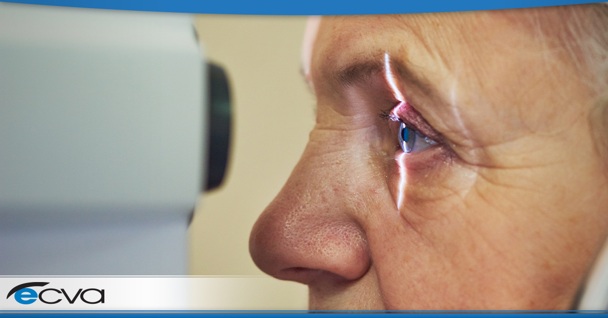
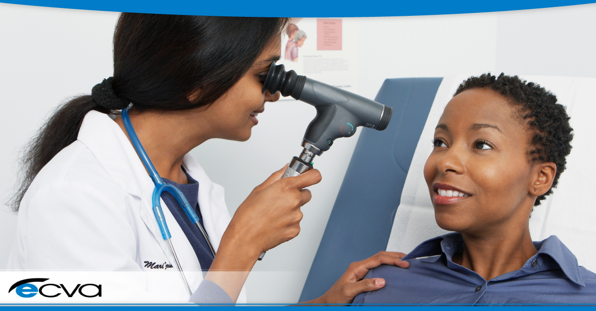
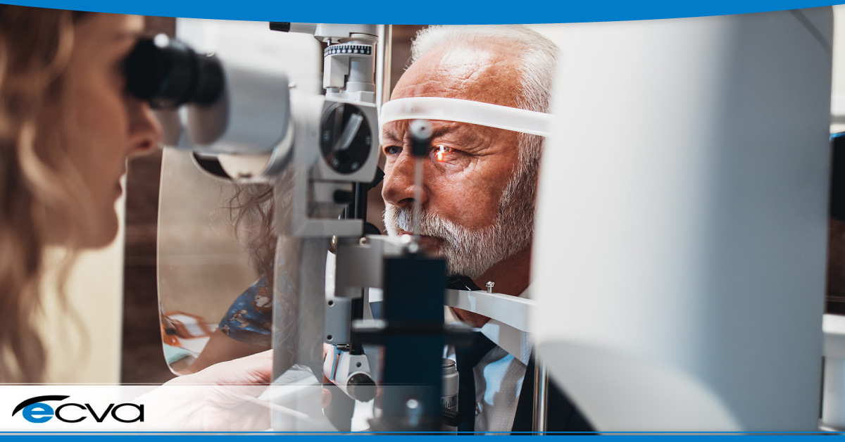
Corneal cross-linking is a minimally invasive procedure designed to strengthen the cornea, the eye’s clear, protective outer layer. The essence of CXL lies in its ability to enhance the corneal fibers’ bonds, thereby increasing the cornea’s structural integrity. This is achieved by applying riboflavin (vitamin B2) drops to the eye, which are then activated by ultraviolet (UV) light. The interaction between the riboflavin and UV light triggers the formation of new collagen bonds within the cornea, making it more robust and resistant to deformation.  

Who Stands to Benefit?  

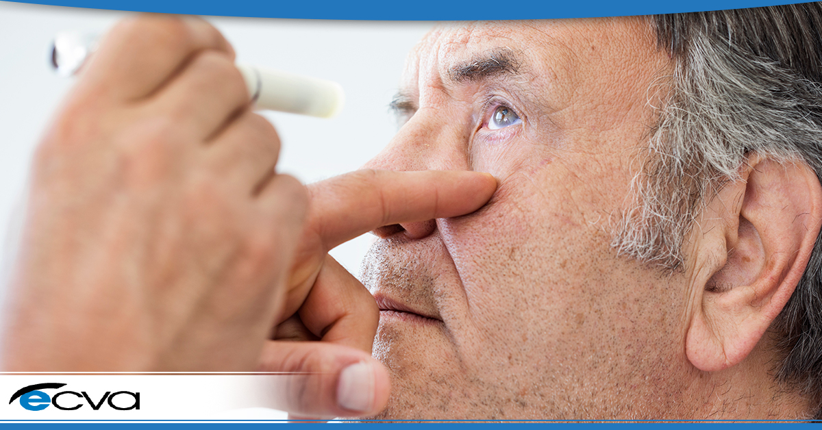
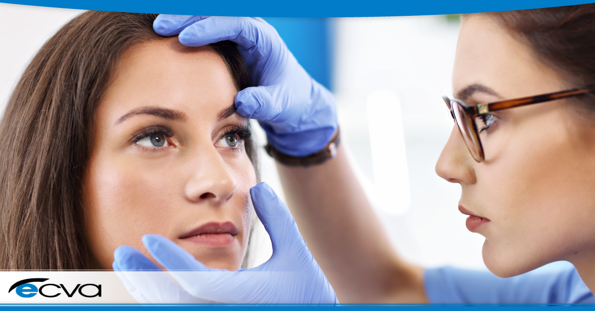
CXL is primarily targeted at individuals suffering from keratoconus, a progressive eye disorder where the cornea thins and gradually bulges outward into a cone shape, causing vision distortion. It is also beneficial for patients with other ectatic disorders or those experiencing corneal weakening due to refractive surgery.  

CXL can prevent further vision deterioration by halting the progression of corneal deformation. It can also avert the need for corneal transplants in advanced cases, allowing patients to avoid a more invasive and higher-risk procedure.  

The Procedure and Recovery Expectations  

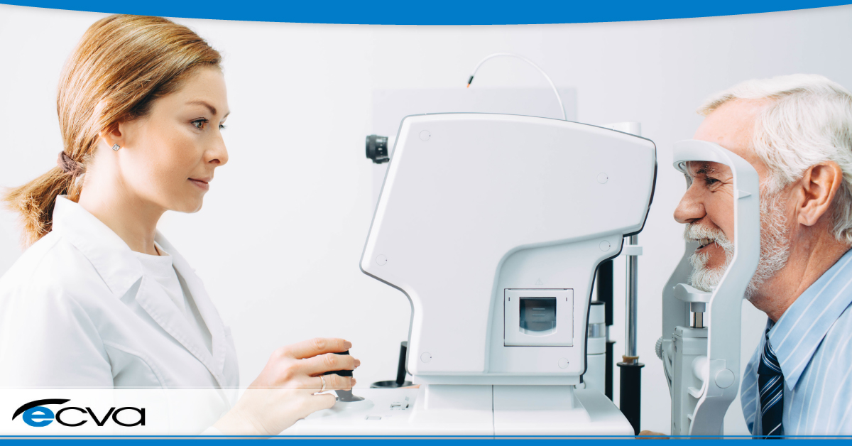
The CXL procedure is performed on an outpatient basis and typically takes about an hour. In most cases, patients should anticipate a relatively straightforward recovery process, although experiences can vary.  

Immediately following the procedure, it’s common to experience some level of discomfort. Additionally, light sensitivity and a foreign body sensation in the eye aren’t uncommon. These symptoms are usually transient. Plus, they can typically be managed with medications prescribed by the treating ophthalmologist.  

Most individuals can return to normal activities within a few days, but it’s crucial to meticulously adhere to post-procedure care instructions. This includes wearing an eye patch or protective shield as advised, avoiding rubbing the eyes, and attending follow-up appointments to monitor healing and corneal stabilization.  

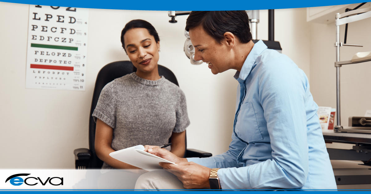
Ultimately, corneal cross-linking represents a beacon of hope for individuals grappling with keratoconus and similar corneal conditions. Its ability to fortify the cornea and arrest the progression of diseases makes it a pivotal treatment in the field of eye health. As with any medical procedure, patient education and setting realistic expectations are key to a successful outcome. Those considering CXL should consult with a qualified ophthalmologist to discuss their suitability for the procedure and to gain a comprehensive understanding of the recovery process.  

Find The Best Cornea Specialist in Buffalo 

At ECVA, we’re dedicated to guiding our patients toward the best possible outcomes. Whether you’re exploring the potential of corneal cross-linking or seeking comprehensive eye care, our team is here to support you every step of the way. Don’t let uncertainty cloud your vision – schedule an appointment at your nearest ECVA clinic today. 

Combined Cataract and Glaucoma Surgery: What to Expect

Both cataracts and glaucoma can rob a person of their vision, and the conditions can occur concurrently. For patients with cataracts and glaucoma, combined cataract and glaucoma surgery is often enticing. During the procedure, your eye care provider addresses both conditions during the surgical procedure, and there are several benefits to the approach.  

If you’re wondering whether combined cataract and glaucoma surgery is the best choice for you and want to know more about what to expect from the procedure, here’s what you need to know.  

The Benefits of Combined Cataract and Glaucoma Surgery  

Combining cataract and glaucoma surgery often significantly improves overall vision quality. The removal of the cloudy lens during cataract surgery, coupled with glaucoma management, enhances visual clarity and sharpness, which makes handling everyday tasks and work activities easier.  

Additionally, a key advantage of opting for combined surgery is patients usually experience a streamlined recovery process. There’s one recovery period instead of two, reducing overall downtime dramatically when compared to having two separate surgeries.  

Finally, combining cataract and glaucoma surgeries is typically more cost-effective than doing procedures separately. Patients often save on total facility fees and other expenses when performing both surgeries at the same time, allowing them to reduce the overall cost of treatment.  

What to Expect from Combined Cataract and Glaucoma Surgery

If you’re considering combined cataract and glaucoma surgery, the first step is typically a comprehensive exam. That allows your eye care provider to evaluate the state of both conditions, confirm you’re a suitable candidate for the procedures, and develop an appropriate treatment plan prior to surgery.  

Once the plan is in place, your eye care provider will discuss what occurs during the cataract removal and glaucoma treatment. For the latter, this usually involves explaining the chosen minimally invasive glaucoma surgery (MIGS) selected or an overview of the available options if more than one MIGS is reasonable.  

During the conversation, your eye care provider will also review the benefits and risks of the combined surgery. The goal is to ensure you’re well-informed, allowing you to decide your best choice is moving forward.  

Once it’s time for your surgery, sedation and local anesthetic are administered to keep you relaxed and to numb the eye. That ensures your comfort during the surgery and can make general anesthesia unnecessary, which reduces overall risk.  

After the surgery, you’ll need to follow the post-surgical recovery plan created and outlined by your provider to support optimal healing. This can include taking specific prescription medications, avoiding strenuous activity, and more, as well as attending scheduled follow-up appointments.  

Are You a Candidate for Combined Cataract and Glaucoma Surgery? 

If you’re interested in combined cataract and glaucoma surgery and want to find out if you’re a suitable candidate, you’ll need to start the process by making an appointment with your eye care provider. Then, they can determine if the procedure is a good treatment choice.  

WNY Best Eye Surgeons

If you’re considering combined cataract and glaucoma surgery, our experienced team is here to provide guidance. Explore the possibilities and take a step towards clearer vision by scheduling an appointment at one of our four convenient ECVA clinic locations in the WNY region. 

Diabetic Eye Disease and Vitreoretinal Surgery: Navigating Treatment Options

Diabetic eye disease like diabetic retinopathy can permanently alter a person’s vision, leading to vision loss or, in severe cases, blindness. With diabetic retinopathy, the blood vessels in the eye weaken. At times, that can lead vessels to bulge and leak, and the latter can cause fluids to enter various parts of the eye, causing retinal tissue swelling. As those tissues are impacted, visual acuity changes, and any damage caused is potentially irreversible.  

However, treating diabetic eye disease can slow or prevent future eye changes, allowing a person to limit their vision loss. One such option is vitreoretinal surgery, also known as vitrectomy, but different paths are also available.  

Here’s a look at vitreoretinal surgery and a few other treatments for diabetic eye disease, as well as what you can do to navigate your treatment options.  

Vitreoretinal Surgery for Diabetic Retinopathy 

Vitreoretinal surgery is a procedure that’s often used in the treatment of advanced diabetic retinopathy. While it isn’t a cure for diabetic retinopathy, it can slow its progression and may reverse some of its impact, causing your vision to possibly improve, stabilize, or avoid significant ongoing losses.  

With a vitrectomy, your eye care provider addresses vitreous gel clouding that can occur when blood vessels leak by removing impacted tissue and replacing it with a clear artificial substitute. In many cases, replacing the cloudy vitreous gel with a clear artificial solution leads to significant vision improvement.  

During the procedure, your eye care provider will also remove built-up scar tissue that’s developed in the eye. Scar tissue can pull on the retina, leading to visual distortions and increasing the chances of a retinal detachment. As a result, scar tissue removal can lead to vision improvements and reduce the risk of retinal detachment.  

Other Treatments for Diabetic Retinopathy

Generally, vitreoretinal surgery is reserved for advanced diabetic retinopathy cases. Other treatment options may provide positive results if your case isn’t that severe. For example, blood sugar management could prevent further damage, which may make additional treatment unnecessary.  

For more severe cases, medication injections involving vascular endothelial growth factor inhibitors can help prevent abnormal blood vessel development that can occur with diabetic retinopathy and reduce fluid buildup. Focal laser or scatter laser treatment – also known as photocoagulation – can also slow or stop blood vessel leakage or shrink abnormal vessels, making them a viable option in some cases.  

How to Navigate Your Treatment Options

Selecting a treatment option after being diagnosed with diabetic retinopathy is often intimidating. Fortunately, by working with your eye care provider, you can receive critical guidance and support that makes selecting an appropriate approach easier. 

Your eye care provider can look at the details of your situation, identifying treatment options that are both viable and have the highest chances of making a positive impact. They can also answer questions to help you understand the recommendations, ensuring you can find the best treatment option for you.  

At ECVA, the safety and health of our patients’ eyes are our priority. If you’ve been diagnosed with diabetic retinopathy, are concerned you may develop diabetic eye disease, or simply haven’t visited your eye care provider in the past year, the ECVA team is here to help. Schedule an appointment at your closest ECVA clinic today.  

The Role of Genetics in Eye Health: Understanding Inherited Eye Conditions

Some eye conditions are related to specific genetic markers. As a result, these eye conditions can be inherited. By understanding what conditions have genetic factors, it’s easier to anticipate your risk of developing them. In turn, it can help you take a more proactive approach to your eye health, which can make a significant difference in overall outcomes.

Here is a look at some common eye conditions that can be linked to genetics.

Myopia and Hyperopia

Both myopia (nearsightedness) and hyperopia (farsightedness) can have a genetic component. Anyone with a first-degree relative with either condition is significantly more likely to develop it than individuals without family members with the condition.

Research involving myopia in children also demonstrates that specific demographics are more likely to develop the condition. For example, one study showed the prevalence is far higher in South Asian children, coming in at nine times greater than among white Europeans.

Glaucoma

Glaucoma is the most common cause of blindness worldwide, as the condition can permanently damage the optic nerve due to increased pressure in the eye. Having a family history of glaucoma puts you at far greater risk, making you four to nine times more likely to develop the condition.

Additionally, people of Hispanic descent are at greater risk than those of European descent. Individuals of Asian descent have a heightened risk of angle-closure glaucoma than other demographics. Glaucoma is also up to eight times more prevalent in the African-American community than in the Caucasian community.

Age-Related Macular Degeneration

Age-related macular degeneration (AMD) – an eye condition that leads to a damaged macula, which causes a reduction in central vision – also seemingly has a genetic component. According to research, an estimated 20 percent of patients with AMD have a family member with the condition, and having a family history makes you four times more likely to develop AMD.

Studies also suggest that Caucasians are more likely to develop AMD than African Americans. Data on whether individuals of Hispanic descent are more or less likely to develop AMD than Caucasians is conflicting, with some reports showing a higher occurrence while others a lower one.

Cataracts

While cataracts are common as people age, specific occurrences of the condition have genetic components. Congenital or childhood bilateral cataracts autosomal dominant inheritance was present among 44 percent of cases examined in one particular study.

Specific ethnic heritages are also at greater risk of developing cataracts in general. For example, African Americans and Latinx Americans are at greater risk than Caucasian Americans.

While a genetic predisposition to specific eye conditions is often concerning, being proactive makes a difference. Proper care and monitoring can lead to an earlier diagnosis, allowing for treatment plans that can slow disease progression and preserve eye health and vision.

At ECVA, the safety and health of our patients’ eyes are our priority. If you’re concerned about whether you’re developing an eye condition with a genetic component or simply haven’t visited your eye care provider in the past year, the ECVA team is here to help. Schedule an appointment at your closest ECVA clinic today.

Common Age-Related Eye Problems

Vision plays a crucial role in everyone’s daily lives. But over time, age-related eye problems may arise, affecting a person’s ability to see clearly and perform common tasks. If you’d like to know about some common age-related eye problems, their causes, symptoms, and potential management strategies, here’s what you need to know. 

Presbyopia 

Presbyopia is one of the most common age-related eye problems. This condition is the result of the loss of elasticity in the lens, making it difficult to focus on nearby objects. People with presbyopia may experience eyestrain, headaches, and might start holding reading materials away from them to improve clarity. Fortunately, presbyopia can be easily managed with reading glasses, bifocals, or multifocal contact lenses. 

Cataracts 

Cataracts are another prevalent eye problem among older adults. They occur when the natural lens of the eye becomes cloudy, leading to blurry vision, faded colors, and increased sensitivity to glare. 

Although cataracts are a natural part of aging, certain risk factors can increase the likelihood that they’ll occur. For example, smoking, excessive sunlight exposure, and diabetes can accelerate the development of cataracts. 

In many cases, cataracts can be treated with surgery. The procedure involves replacing the cloudy lens with an artificial one, and the artificial lens restores clear vision. However, if symptoms are mild, using brighter lighting, wearing anti-glare glasses or sunglasses, or other less invasive options may work well. 

Age-Related Macular Degeneration 

Age-related macular degeneration (AMD) is a progressive eye condition that impacts the macula, the part of the eye responsible for central vision. When a person has AMD, it typically causes a gradual loss of vision in the middle of the visual field. That makes it challenging to read, recognize faces, or perform tasks that require fine visual acuity. 

While the exact cause of AMD isn’t known, specific factors can play a role. For example, genetics, smoking, obesity, and high blood pressure can increase the risk. 

Although there is no cure for AMD, early detection and lifestyle modifications, such as a healthy diet and protecting your eyes from harmful UV rays, can help slow its progression. 

Glaucoma 

Glaucoma is a set of eye conditions that harm the optic nerve, typically due to increased pressure in the eye, which leads to damage. The most widely experienced type of glaucoma is primary open-angle glaucoma. In most cases, it develops slowly and painlessly, gradually affecting peripheral vision. 

If left untreated, glaucoma can lead to permanent vision loss. Regular eye examinations, along with medications, eye drops, or surgical interventions, can help manage glaucoma and prevent further damage. 

Dry Eye Syndrome 

Dry eye syndrome is when the eyes don’t produce enough tears or when a person’s tears evaporate too quickly. Tear production usually decreases as a person ages, leading to dryness, irritation, and a gritty sensation in the eyes. 

Environmental factors, medications, and specific underlying health conditions can exacerbate dry eye symptoms. Artificial tears, lifestyle adjustments – such as blinking more frequently and using a humidifier – and prescription medications can help manage dry eye syndrome effectively. 

At ECVA, the safety and health of our patients’ eyes are our priority. If you’re concerned about age-related eye problems or simply haven’t seen your eye care provider in the past year, the ECVA team is here to help. Schedule an appointment at your closest ECVA clinic today. 

Types of Retinal Detachment

Retinal detachments dramatically alter your vision and can lead to permanent changes that cost you your eyesight. While many people are at least somewhat aware of the condition, many patients aren’t overly familiar with the different types of retinal detachment.  

Technically, all retinal detachments involve the retina moving away from the back of the eye, leading to visual distortions, blind spots, and other symptoms. However, how they occur varies. Retinal detachments are separated into three categories: exudative, rhegmatogenous, and tractional. Each represents a different cause for a retinal detachment.  

Here is an overview of the three types of retinal detachment.  

Exudative  

Exudative retinal detachment happens when fluid begins building up behind the retina. As the fluid level rises, it puts pressure on the retina, eventually causing it to tear away from the back of the eye.  

In most cases, exudative retinal detachments are caused by swelling in the back of the eye or blood vessels in or near the back of the eye leaking. Causes of swelling or leaking vessels can include injuries, age-related macular degeneration, disease-related inflammation, and tumors. Individuals with Coats disease are also at risk.  

Rhegmatogenous  

Rhegmatogenous retinal detachments are the most prevalent kind of retinal detachment. These occur due to a small break or tear in the retina that allows the vitreous – a gel-like fluid in the eye – to flow behind the retina. As the vitreous positions itself behind the retina, it pushes against the retina. When the pressure is high enough, a detachment occurs.  

Most cases of rhegmatogenous retinal detachment are related to aging. As you get older, the vitreous can shrink or experience texture changes. At times, those changes pull on the retina, resulting in a tear. However, other situations – including eye surgeries, eye injuries, and nearsightedness – can also cause this type of retinal detachment.  

Tractional  

With tractional retinal detachments, the cause is typically scar tissue. As the scar tissue develops, it can tug at the retina, essentially pulling it away from the back of the eye.  

Generally, tractional retinal detachment is most commonly associated with diabetic retinopathy, an eye condition that individuals with diabetes may develop. Diabetic retinopathy leads to blood vessel damage, which can cause scarring. However, eye infections and other diseases can also cause retinal detachment. The same is true of eye swelling.  

Regardless of the cause, retinal detachment is a severe medical condition that requires immediate treatment. With quick treatment, vision loss can be minimized and even potentially reversible in some cases. As a result, anyone experiencing signs of retinal detachment – such as a sudden increase in floaters, bright light flashes, blurred vision, or darkening vision, among others – should seek emergency medical care immediately.  

At ECVA, the safety and health of our patients’ eyes are our priority. If you’re experiencing symptoms of a potential retinal attachment, require care relating to a past retinal detachment, or simply haven’t seen your eye care provider in the past year, the ECVA team is here to help. Schedule an appointment at your closest ECVA clinic today.  

What Causes Tunnel Vision?

Tunnel vision is a highly disruptive condition, making it difficult or impossible to see objects that aren’t positioned directly in front of you. Without your peripheral vision, navigating the world becomes riskier, as you can’t clearly identify objects to your side. As a result, it’s critical to understand what causes tunnel vision and whether it’s correctable. Here’s what you need to know. 

What Is Tunnel Vision? 

Technically referred to as peripheral vision loss, tunnel vision is functionally the loss or diminishment of your peripheral vision. When it occurs, objects that aren’t directly in front of you become unclear or impossible to see. Essentially, your field of vision shrinks, leaving only the central part intact. 

What Causes Tunnel Vision? 

There are several potential causes of tunnel vision. In some cases, medical conditions impacting the brain can lead to the condition. Issues with blood vessels or nerves connected to your eyes or vision centers may also cause tunnel vision. 

Additionally, there are eye conditions that may lead to tunnel vision. Diabetic retinopathy, glaucoma, retinitis pigmentosa, retinal detachment, retinal vein occlusion, and optic neuritis are all eye diseases that have the potential to cause tunnel vision. 

In some cases, migraines may lead to peripheral vision loss. The same is true of hypertension, concussions, and strokes. 

Is Tunnel Vision Serious? 

Any change to your vision is serious. With tunnel vision, the loss of your peripheral vision impacts your daily life. Along with altering what you can and can’t see clearly, it can lead to headaches, dizziness, and other uncomfortable symptoms, which are often highly disruptive. 

Additionally, some conditions require immediate treatment to have the best chance of restoring your vision. Since that’s the case, treating any occurrence of peripheral vision loss as a serious medical event and seeking care immediately is wise. 

Finally, some causes of tunnel vision are also life-threatening. Conditions like strokes can lead to peripheral vision loss, though there may be other damage that isn’t causing immediately clear symptoms. If treatment is delayed, the stroke can cause more damage to the brain, which can lead to catastrophic outcomes, including loss of life. 

Can You Recover from Tunnel Vision? 

Whether tunnel vision is temporary or permanent depends on the cause of the peripheral vision loss. For example, tunnel vision relating to migraines often clears once the migraine subsides. If retinal detachment is the cause, tunnel vision isn’t recoverable unless the proper treatment option is available and pursued quickly. With diabetic retinopathy, stroke, or glaucoma, the peripheral vision loss is often permanent. 

As a result, it’s critical to see a medical professional or your eye care provider right away if you’re experiencing tunnel vision. They can determine the cause and recommend treatments that may restore your vision, depending on what underlying condition is responsible. Additionally, some causes of peripheral vision loss are life-threatening, making immediate care essential. 

At ECVA, the safety and health of our patients’ eyes are our priority. If you’re experiencing tunnel vision or simply haven’t seen your eye care provider in the past year, the ECVA team is here to help. Schedule an appointment at your closest ECVA clinic today. 

Family looking at eyeglasses

Questions to Ask Your Doctor About AMD

Receiving a diagnosis of age-related macular degeneration (AMD) is often startling and concerning, particularly if you aren’t overly familiar with AMD. As a result, it’s wise to prepare questions that you can ask your ophthalmologist after being referred for an appointment. If you aren’t sure what to ask, here are a few questions that will help you understand the diagnosis and get the conversational ball rolling. 

Is It Wet or Dry AMD, and Is It Affecting One or Both Eyes? 

AMD comes in two primary forms, wet and dry. Each type has a unique cause and requires different treatments to manage. As a result, it’s critical to learn which kind of AMD you have early in the process. 

Additionally, finding out if it’s affecting one or both eyes is wise. Either is a possibility, so learning whether both eyes are impacted is essential. 

What Stage Is My AMD? 

Knowing the stage of AMD lets you know how advanced the condition is, and it may make it easier to determine which treatment options are worth pursuing at that time. By asking your ophthalmologist whether you’re in an early, intermediate, or advanced stage, you’ll have a better understanding of your current diagnosis. 

Is My AMD Likely to Progress? 

When, how, or if a condition progresses depends on multiple factors. With this question, you can find out how quickly your AMD may advance, allowing you to determine whether various treatment options make sense at this time. 

Will Any Lifestyle Changes Slow Progression? 

In some cases, lifestyle changes – like altering your diet, adjusting your exercise schedule, or taking supplements – can impact the progression of specific eye conditions. However, whether any changes are right for you depends on many factors. For example, whether there are other medical conditions you have that would be negatively impacted by an adjustment is something you need to discuss with your care team before making lifestyle changes. 

Should My Blood Relatives Get Checked for AMD? 

Genetics can play a role in some eye conditions, including whether someone is potentially at greater risk of developing AMD. By asking your doctor this question, you can find out if your blood relatives – such as siblings or children – may need more frequent screenings to ensure the health of their eyes. 

How Frequently Should I Schedule Follow-Ups with My Eye Care Provider? 

With conditions like AMD, it’s wise to schedule follow-up appointments with your eye care provider to monitor how your AMD progresses and whether various treatments are effective. Speak with your ophthalmologist to determine an ideal schedule and prioritize those appointments. That ensures your eye care provider can adjust the treatment plan quickly if necessary, increasing the odds that your vision is preserved as much as possible. 

At ECVA, the safety and health of our patients’ eyes are our priority. If you were recently diagnosed with AMD and want guidance on treatment options, our board-certified ophthalmologists are here to help. Schedule an appointment at your closest ECVA clinic today. 

Ways to Take Better Care of Your Eyes in 2023

Caring for your eyes is an excellent way to help keep them healthy. Specific eye care habits can reduce your odds of developing specific conditions, while others can keep your eyes comfortable during changing environmental conditions. 

Fortunately, creating an effective eye care routine is typically far simpler than most people expect. If you are prioritizing your health this new year, here are a few simple ways to care for your eyes every day. 

Wash Your Hands Regularly (and Avoid Touching Your Eyes) 

Regular hand washing helps remove bacteria, dust, and dirt, reducing the odds of transfer if you contact your eye area. By also avoiding touching and rubbing your eyes, you make it even less likely that any substance or bacteria will infect your eyes. Plus, not rubbing can reduce irritation if anything is in your eye, making injuries less likely, too. 

Adopt Healthy Eating Habits 

Eating a healthy, balanced diet and drinking enough water can improve your eye health. Remaining hydrated can prevent your eyes from getting dry and irritated. Nutrients like omega-3 fatty acids, vitamin A, vitamin C, vitamin E, lycopene, lutein, beta-carotene, and others also help ensures your eyes have what they need to stay healthy. 

Quit Smoking 

Smoking isn’t just harmful to your lungs; it also negatively impacts your eyes. Smokers are more at risk of developing certain conditions, including cataracts, age-related macular degeneration, and others. Smoking also leads to optic nerve damage that can reduce your visual acuity over time, even if it doesn’t lead to another condition. By quitting, you reduce your risk significantly, so it’s worth doing. 

Where UV-Blocking Glasses 

Ultraviolet (UV) rays can damage your eyes in several ways. Corneas are susceptible to sunburns which, along with being uncomfortable, can potentially lead to vision issues. Exposure to UV rays is also associated with conditions like photokeratitis and age-related macular degeneration. 

By wearing UV-blocking sunglasses or getting UV-blocking coatings on regular glasses lenses, you can filter out harmful UV light. Coupling those with hats or visors can also make the approach more effective. 

Use Screens Correctly 

Digital devices and screens can lead to a variety of eye issues. Improper distances, excessive glare, or extended durations classically lead to problems like eye strain and dry eye. 

When you’re using screens, keep the brightness and angle appropriate. For computer screens, that generally means keeping the monitor about 20 degrees below eye level and the distance close to arm’s length. For any screen, make sure you look away every 20 minutes and focus on a spot that’s about 20 feet away from your position. It’s also wise to take breaks regularly, giving you a chance to move around and rest your eyes before returning. 

At ECVA, the safety and health of our patients’ eyes are our priority. We recommend that everyone get their eyes checked annually by an eye doctor to ensure eye health and early detection of any problems. Schedule an appointment at your closest ECVA clinic today, we’d love to see your family for a checkup. 

What Is Thyroid Eye Disease?

hyroid eye disease (TED) is an autoimmune condition that impacts eye tissues. Typically, TED leads to inflammation, swelling, and damage to the muscles, connective tissues, and fatty tissues in the eye area. 

Since TED can cause permanent, lasting damage, understanding what it does, who’s at risk, early systems, and why prompt treatment is critical is essential. Here’s a closer look at thyroid eye disease. 

What Thyroid Eye Disease Does to Eye Tissues 

As commonly occurs with autoimmune conditions, the person’s immune system is essentially attacking their own body. With TED, the eye tissues are targeted by the immune system. 

TED occurs in two phases. First, there’s the active phase, which is also known as the inflammatory phase. This period can last up to three years, and it’s generally marked by ongoing inflammation and inflammation-related symptoms and damage. Second, there’s the stable phase, where inflammation subsides. 

TED can cause lead to other conditions, such as secondary glaucoma. Regardless of whether that occurs, the damage created by TED is potentially impactful. 

Who Is at Risk of Thyroid Eye Disease? 

In many cases, TED is connected to Graves disease, an autoimmune condition that affects the skin, thyroid, and eyes. Graves disease can lead to hyperthyroidism or hypothyroidism, both of which can trigger TED. 

Hyperthyroidism or hypothyroidism not related to Graves disease may also result in TED. However, TED can occur without Graves disease or thyroid hormone abnormalities. Another risk factor is low levels of selenium in your blood. 

The Early Symptoms of Thyroid Eye Disease 

There are many symptoms associated with TED. While one of the most pronounced is proptosis (bulging eyes), dry eyes, watery eyes, irritation caused by a gritty feeling, redness, and double vision may occur before there are obvious physical changes. 

Vision changes can occur, as well as pain with eye movements or discomfort behind the eyes. Trouble closing your eyes fully is also a potential symptom and may lead to a corneal ulcer. 

In some cases, symptoms may only affect one eye. However, they can also occur in both. 

Why Regular Eye Appointments Are Crucial 

Scheduling regular eye appointments is critical regardless of whether you currently have TED. Your eye care provider can look for signs and symptoms that may indicate TED or conditions that may lead to thyroid eye disease. 

If you have TED, frequent appointments allow your eye care provider to monitor your condition and provide treatment options. This may include over-the-counter options to relieve irritation or dryness and reduce inflammation, selenium supplements if the levels in your blood are low, or prescription medications. 

Your eye care provider may also recommend certain home remedies or lifestyle changes. For example, using cool compresses to reduce swelling and discomfort may be part of the plan. Keeping your head higher when lying down, wearing sunglasses when in well-lit spaces or outdoors, eyelid taping, and similar steps may also be included. 

In some cases, surgical treatments might be on the table. This can include eyelid, eye muscle, or orbital decompression procedures. Radiation therapy is a potential option for combating inflammation, as well. 

At ECVA, the safety and health of our patients’ eyes are our priority. If you are concerned about thyroid eye disease or simply haven’t seen your eye care provider in the past year, the ECVA team is here to help. Schedule an appointment at your closest ECVA clinic today.